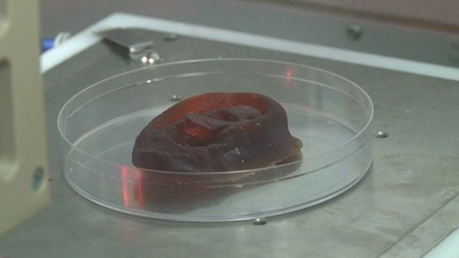
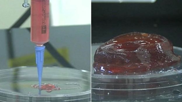
 It is hoped the grown cartilage will be used on humans in about three years
Patients needing surgery to reconstruct body parts such as noses and ears could soon have treatment using cartilage which has been grown in a lab.
The process involves growing someone's cells in an incubator and then mixing them with a liquid which is 3D printed into the jelly-like shape needed.
It is then put back in an incubator to grow again until it is ready.
Researchers in Swansea hope to be among the first in the world to start using it on humans within three years.
"In simple terms, we're trying to grow new tissue using human cells," said Prof Iain Whitaker, consultant plastic surgeon at the Welsh Centre for Burns and Plastic Surgery at Morriston Hospital.
"We're trialling using 3D printing which is a very exciting potential modality to make these relatively complex structures.
"Most people have heard a lot about 3D printing and that started with traditional 3D printing using plastics and metals.

"That has now developed so we can consider printing biological tissue called 3D bio-printing, which is very different.
"We're trying to print biological structures using human cells, and provide the right environment and the right timing so it can grow into tissue that we can eventually put into a human.
"It would be to reconstruct lost body parts such as part of the nose or the ear and ultimately large body parts including bone, muscle and vessels."
The team of surgeons are working with scientists and engineers who have built a 3D printer specifically for this work.
Prof Whitaker, who is also the chairman of plastic and reconstructive surgery at Swansea University's medical school, said the project started in 2012 but research in the field has been going on for more than 20 years.
He said the work would have to be tested on animals and go through an ethics process before being used on humans.
"The good news in the future is, if our research is successful, within two months you'd be able to recreate a body part which was not there without having to resort to taking it from another part of the body which would cause another defect or scar elsewhere," he added.
How the process works
 ABMU
ABMU- Cells are taken from a tiny sample of cartilage during the initial operation and grown in an incubator over several weeks
- The shape of the missing body part is scanned and fed into a computer
- It is then 3D printed using a special liquid formula combined with the live cells to form the jelly-like structure
- Reagents are added to strengthen the structure
- It is put into an incubator with a flow of nutrients to supply the cells with food so they can grow and produce their own cartilage
- The structure will then be tested to see if it is strong enough to be eventually implanted into patients


No comments:
Post a Comment